

鈦和不銹鋼等植入物通常覆蓋羥基磷灰石塗層,以欺騙身體並降低植入物排斥率。
羥基磷灰石可使用於骨空隙或缺陷的,透過將材料的粉末、塊或顆粒放置在受影響的骨骼區域來進行修復。
羥基磷灰石的生物活性可以促進骨骼生長並修復缺損,並且可以替代骨移植。
如果不使用羥基磷灰石,它通常會導致癒合時間比觀察到的要長。
受損骨骼使用nanoXIM填充過程
用 nanoXIM 羥基磷灰石和磷酸三鈣生產的骨替代品具有生物相容性、骨傳導與骨激發性,
其納米結構與人體骨骼具有高度的相似性,是最有前景的骨再生合成材料。
一旦放置在受損的骨骼區域,nanoXIM 很容易與宿主組織結合併整合,
從而誘導天然骨骼的形成和生, 最後組織將被完全重建,材料將被原生骨替代。

應用
-
-
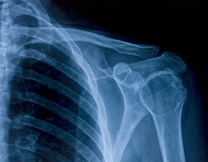
- 儘管骨骼具有良好的自愈能力,但較嚴重的骨損傷缺損是無法自愈的。
- 合成材料能夠克服骨缺損常用治療方法(自體骨移植)的缺點。
- 使用合成材料代表不需要從髂骨稜取骨,減少併發症的發生、後續相關手術。
- 無限可用性
-

推薦用於製造骨移植替代品,例如:骨再生的注射劑和硬組織植入物。
nanoXIM HAp 糊狀物對硬組織具有高親和力,可與宿主組織建立化學鍵。
- 羥基磷灰石相純度 (100%)
- 羥基磷灰石納米粒子(< 50 nm)
- 快速促進骨再生和早期血管形成
- 促進蛋白質吸附和成骨細胞粘附
- 增強成骨細胞功能
- 與人體相容性高
- 材料可以在癒合過程中被新骨替代、吸收
- 膏狀具有最佳的缺陷填充能力
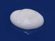 |  | 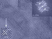 |
| 膏狀 | 高解析 | 電子晶體學圖像 |
Stem Cell Applications of Nanohydroxyapatite納米羥基磷灰石的干細胞應用
nanoXIM HAp pastes are nano-hydroxyapatite water based pastes specially recommended to manufacture bone graft substitutes such as injectables for bone regeneration and implants for hard tissues.
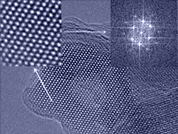
The hydroxyapatite nanoparticles comprised in these products form a perfectly aligned structure of nanocrystals.
Due to the similarity between nano-hydroxyapatite and mineralized bone, nanoXIM HAp pastes have a high affinity to hard tissues, establishing chemical bonds with the host tissue.

 Benefits
| Promotes fast bone regeneration and an early vascularization due to their osteoconductive and osteostimulative properties | |
| Encourages protein adsorption and osteoblast adhesion | |
| Enhances osteoblast functions | |
| Biocompatible material | |
| Resorbable material replaced by new bone during the healing process | |
| Optimal defect filling capacity due to pasty consistency |
 Features
| Hydroxyapatite phase purity (100%) | |
| Hydroxyapatite nanoparticles (< 50 nm) | |
| High surface area | |
| Synthetic material |

 Technical Data Sheet
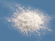
nanoXIM•HAp100 is a series of synthetic nano-hydroxyapatite aqueous pastes, manufactured and supplied in two different concentrations, 15 and 30 wt%.
These products comprise nano-hydroxyapatite particles with typical particle size below 50 nm in a rod-like shape (typically 30-40 nm length and 5-10 nm width) suspended in pure water.
| Reference | Hydroxyapatite (wt%) | |
| nanoXIM•HAp102 | 15±1.0 | ADD |
| nanoXIM•HAp103 | 30±3.0 | ADD |
Disclaimer: nanoXIM products are supplied in bulk and in non-sterile form.
 nanoXIM.HAp Paste |  High Resolution TEM of |  Electron crystallography image |
推薦用於製造生物相容性骨移植替代品(例如:骨再生的多孔顆粒和支架)和 3D 打印植入物。
nanoXIM HAp_粉末可以在一段時間內逐漸分解並被宿主的組織替代。
- 羥基磷灰石相純度 (100%)
- 奈米結構微米級粉末
- 高表面積
- 快速促進骨再生和血管化
- 骨傳導與骨激發性
- 增強成骨細胞功能
- 與人體相容性高
- 符合 ISO 13779 的重金屬和 Ca/P 比
 |  |  |
| 粉末 | 高解析 | 電子晶體學圖像 |
Improving Bone Regeneration Using Nanohydroxyapatite 使用 HAp 改善骨再生
Applications of Nanostructured Hydroxyapatite for Osteogenesis and Angiogenesis 奈米結構HAp在成骨和血管生成中的應用
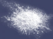
nanoXIM HAp powders are micrometric aggregates of hydroxyapatite nanoparticles.
These products are used in the manufacturing of biocompatible bone graft substitutes (e.g. porous granules and scaffolds for bone regeneration) and 3D printed implants.
Due to the similarity between nano-hydroxyapatite and mineralized bone, nanoXIM HAp powders have a high affinity to hard tissues as they form chemical bonds with the host tissue resulting in an improved biological performance.
Benefits
| Promotes bone regeneration and vascularization | |
| Osteoconductive and osteostimulative | |
| Enhances osteoblast functions | |
| Biocompatible material |
Features
| Pure hydroxyapatite | |
| Micron-sized powders | |
| Narrow particle size distribution | |
| Synthetic material | |
| Complies with heavy metals and Ca/P ratio according to ISO 13779 |
推薦用於製造骨替代的塊狀、顆粒狀和磷酸鈣水泥
nanoXIM TCP_粉末可以在一段時間內逐漸分解並被宿主的組織替代。
- 不含鈣的羥基磷灰石粉末
- 羥基磷灰石相純度 (100%)
- 奈米結構微米級粉末
- 高表面積
- 高孔隙率
- 快速促進骨再生和早期血管形成
- 促進蛋白質吸附和成骨細胞粘附
- 增強成骨細胞功能
- 與人體相容性高
- 材料可以在癒合過程中被新骨替代、吸收
 |  |  |
| 粉末 | 高解析 | 電子晶體學圖像 |
nanoXIM TCP is a calcium deficient hydroxyapatite powder consisting of nanostructured micron-sized aggregates.
This product is used to manufacture blocks, granules and calcium phosphate cements for bone replacement, allowing a gradual biological degradation over a period of time and a progressive replacement by the natural host tissue.
Benefits
| Promotes fast bone regeneration and an early vascularization due to their osteoconductive and osteostimulative properties | |
| Encourages protein adsorption and osteoblast adhesion | |
| Enhances osteoblast functions | |
| Biocompatible material | |
| Resorbable material replaced by new bone during the healing process |
Features
| Pure calcium deficient hydroxyapatite | |
| High surface area | |
| High porosity | |
| Nanostructured micron sized powder | |
| Synthetic material |
Technical Data Sheet
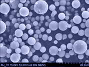
nanoXIM•TCP200 powder is a synthetic calcium phosphate form commonly designated by calcium-deficient hydroxyapatite. Once sintered at 1000ºC (following ISO 13779 procedures), a minimum of 95% β-TCP phase purity is ensured in accordance with ASTM F1088-04ɛ1.
nanoXIM•TCP200 powder is supplied as synthetic nanostructured micron-size particles of 5 μm with a high specific surface area. This feature is achieved in the drying process by spray dryer technique where the nanoparticles in liquid phase are dried as spherical aggregates.
| Reference | Particle size, d50 (μm) | |
| nanoXIM•TCP200 | 5.0±2.0 | ADD |
Disclaimer: nanoXIM products are supplied in bulk and in non-sterile form.
 nanoXIM TCP Powders |  SEM of nanoXIM.TCP Powder |  Electron crystallography image |
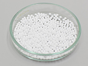
nanoXIM•Granules_球型顆粒劑 球形形狀的顆粒擁有良好的流動性和處理性能、可以形成均勻的粒間孔。
- 羥基磷灰石相純度 (100%)
- 高度均勻的顆粒大小
- 能夠填充不規則形狀的孔洞、缺陷
- 良好的流動性和處理性能
- 由於球形而形成均勻的粒間孔
 | 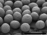 | 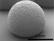 |
| 球型顆粒 | 高解析 G1000 | 高解析 G2000 |
nanoXIM•Granules are spherical granules of synthetic hydroxyapatite available in two different particle ranges (0.5 - 1.0 and 1.0 - 2.0 mm). The high similarity of nano-hydroxyapatite to natural bone results in improved biological performance during bone regeneration.
nanoXIM•Granules with uniform size and spherical shape not only allows good flowability and handling properties, but also the formation of homogeneous intergranular pores.
Benefits
| Biocompatibility | |
| Able to fill irregular shaped hollows/cavities/defects | |
| Good flowability and handling properties | |
| Formation of homogeneous intergranular pores due to spherical shape |
Features
| Highly homogeneous granules size | |
| Fully synthetic | |
| Hydroxyapatite with high phase purity | |
| Available in two particle ranges |
Technical Data Sheet
nanoXIM•Granules are spherical granules composed of aggregates of synthetic nano-hydroxyapatite.
These granules are available in two particle ranges, 0.5 - 1.0 mm and 1.0 - 2.0 mm, so they can be used to create medical devices able to fill different and patient-specific bone defects.
| Reference | Particle range (mm) | |
| nanoXIM•G1000 | 0.5 - 1.0 | ADD |
| nanoXIM•G2000 | 1.0 - 2.0 | ADD |
Disclaimer: nanoXIM•Granules are not finished medical devices, not sterile and are sold in bulk. As they are not finished products, nanoXIM•Granules do not have CE mark or FDA approval and these certifications must be ensured by our clients.
 nanoXIM•Granules |  SEM image of nanoXIM•G1000 |  SEM image of nanoXIM•G2000 |
- Stem Cell Applications of Nanohydroxyapatite
納米羥基磷灰石的干細胞應用
在骨再生過程中,人體中的間充質乾細胞 (HMSC) 發揮重要作用,
它們移動到受傷部位並分化為骨細胞,從而實現再生過程。
考慮到這些細胞的重要性,評估了nanoXIM • HAp102在HMSCs增殖和成骨細胞分化中的生物學性能。

- Improving Bone Regeneration Using Nanohydroxyapatite
使用 HAp 改善骨再生
形成穩定的微血管系統是為了確保骨組織成功再生的必要過程。
在這項研究中,使用nanoXIM•HAp202生產的 3D 顆粒讓血管生成以及成骨。
為此,在nanoXIM•HAp202顆粒上進行了人類真皮微血管內皮細胞 (HDMEC) 和人體間充質乾細胞 (HMSC) 的共培養。

- Applications of Nanostructured Hydroxyapatite for Osteogenesis and Angiogenesis
奈米結構HAp在成骨和血管生成中的應用
在骨再生過程中,骨細胞的活力和增殖是必不可少的。
在這項研究中,使用nanoXIM•HAp202 與用 micro HAp 培養的 MG63 細胞(成骨細胞樣細胞)比較基質上的活力和增殖。
在這兩種情況下,含有nanoXIM•HAp202的生物材料的細胞活力和增殖都更高。

Y. Liu, A. Nadeem, S. Sebastian, M.A. Olsson, S.N. Wai, E. Styring, J. Engellau, H. Isaksson, M. Tägil, L. Lidgren, D.B. Raina, “Bone mineral: A trojan horse for bone cancers. Efficient mitochondria targeted delivery and tumor eradication with nano hydroxyapatite containing doxorubicin”, Materials Today Bio, 14, 100227 (2022).
K.K. Moncal, R.S.T. Aydın, K.P. Godzik, T.M. Acri, D.N. Heo, E. Rizk, H. Wee, G.S. Lewis, A.K. Salem, I.T. Ozbolat, “Controlled Co-delivery of pPDGF-B and pBMP-2 from intraoperatively bioprinted bone constructs improves the repair of calvarial defects in rats,” Biomaterials, 281, 121333, (2022).
D.V. Blase, R.G. Dricot, J.F. Lasserre, S. Toma, M.C. Brecx, “Combination of a Hydraulic Device and Nanohydroxylapatite Paste for Minimally Invasive Transcrestal Sinus Floor Elevation: Procedure and 4-Year Results.”, The International Journal of Oral & Maxillofacial Implants, 36(3), p. 587 (2021).
P. Melo, G. Montalbano, S. Fiorilli, C. Vitale-Brovarone, “3D Printing in Alginic Acid Bath of In-Situ Crosslinked Collagen Composite Scaffolds”. Materials, 14(21), 6720 (2021).
N. Ribeiro, A. Sousa, C. Cunha-Reis, A.L. Oliveira, P.L. Granja, F.J. Monteiro, S.R. Sousa, “New prospects in skin regeneration and repair using nanophased hydroxyapatite embedded in collagen nanofibers”, Nanomedicine: Nanotechnology, Biology and Medicine, 33, 102353 (2021).
P. Melo, R. Naseem, I. Corvaglia, G. Montalbano, C Pontremoli, A. Azevedo, P. Quadros, P. Gentile, A.M. Ferreira, K. Dalgarno, C. Vitale-Brovarone, S. Fiorilli, “Processing of Sr2+ Containing Poly L-Lactic Acid-Based Hybrid Composites for Additive Manufacturing of Bone Scaffolds”, Front. Mater. 7:601645. doi: 10.3389/fmats.2020.601645 (2020).
H Yue, Q Zhu, S Dong, Y Zhou, Y Yang, L Cheng, M. Qian, L. Liang, W. Wei, H. Wang “A Nanopile Interlocking Separator Coating towards Uniform Li Deposition of the Li Metal Anodes” ACS Appl. Mater. Interfaces, doi:10.1021/acsami.0c08776 (2020).
R.V. Pinto, P.S. Gomes, M.H. Fernandes, M.E.V. Costa, M.M. Almeida, “Glutaraldehyde-crosslinking chitosan scaffolds reinforced with calcium phosphate spray-dried granules for bone tissue applications”, Materials Science & Engineering C, 109, 110557, doi:10.1016/j.msec.2019.110557 (2020) .
I. Corvaglia, “Design and optimization of hybrid formulations based on PLLA and inorganic phases for 3D printing of bone scaffolds”, MSc thesis in Biomedical Engineering, Politecnico di Torino (2020).
L. Siqueira, N. Ribeiro, M.B.A. Paredes, L. Grenho, C. Cunha-Reis, E.S. Trichês, M.H. Fernandes, S.R. Sousa, F.J. Monteiro , “Influence of PLLA/PCL/HA Scaffold Fiber Orientation on Mechanical Properties and Osteoblast Behavior”, Materials, 12, 3879, doi:10.3390/ma12233879 (2019).
R.N. Salaie, A. Besinis, H. Le, C. Tredwin, R.D. Handy, “The biocompatibility of silver and hydroxyapatite coatings on titanium dental implants with human primary osteoblast cells”, Materials Science & Engineering C, doi.org/10.1016/j.msec.2019.110210 (2019).
D. Kołbuk, O. Urbanek, P. Denis, E. Choińska, “Sonochemical coating as an effective method of polymeric nonwovens functionalization”, Journal of Biomedical Materials Research – Part A, p. 65 (2019).
C. Santos, S. Turiel, P.S. Gomes, E. Costa, A. Santos Silva, P. Quadros, J. Duarte, S. Battistuzzo, M.H. Fernandes, “Vascular biosafety of commercial hydroxyapatite particles: discrepancy between blood compatibility assays and endothelial cell behavior”, Journal of Nanobiotechnology, 16(27), doi: 10.1186/s12951-018-0357-y (2018).
G. Ruphuy, T. Weide, J.C.B. Lopes, M.M. Dias, M.F. Barreiro, “Preparation of nano-hydroxyapatite/chitosan aqueous dispersions: from lab scale to continuous production using an innovative static mixer”, Carbohydrate Polymers, 202, p. 20 (2018).
D. Dzhurinskiy, “Bioactive antimicrobial coatings for implantable medical devices formed by plasma electrolytic oxidation”, Metal Forming XXIX (1), p. 65 (2018).
J.D.F. Queiroz, “Avaliação da resposta celular a biomateriais para fins de regeneração óssea”, PhD Thesis in Health Sciences, Universidade Federal do Rio Grande do Norte (2018).
G. Ruphuy, M. Souto-Lopes, D. Paiva, P. Costa, A.E. Rodrigues, F.J. Monteiro, C.L. Salgado, M.H. Fernandes, J.C. Lopes, M.M. Dias, M.F. Barreiro, “Supercritical CO2 assisted process for the production of high-purity and sterile nano-hydroxyapatite/chitosan hybrid scaffolds”, J Biomed Mater Res Part B, DOI: 10.1002/jbm.b.33903 (2017).
Y. Ryabenkova, A. Pinnock, P.A. Quadros, R.L. Goodchild, G. Möbus, A. Crawford, P.V. Hatton, C.A. Miller, “The relationship between particle morphology and rheological properties in injectable nano-hydroxyapatite bone graft substitutes”, Materials Science and Engineering: C, 75, p. 1083, (2017).
V. Hruschka, S. Tangl, Y. Ryabenkova, P. Heimel, D. Barnewitz, G. Möbus, C. Keibl, J. Ferguson, P. Quadros, C. Miller, R. Goodchild, W. Austin, H. Redl, T. Nau, “Comparison of nanoparticular hydroxyapatite pastes of different particle content and size in a novel scapula defect model”, Nature Scientific Reports 7, Article number: 43425; doi: 10.1038/srep43425 (2017).
A. Besinis, S. D. Hadi, H. R. Le, C. Tredwin, R. D. Handy, “Antibacterial activity and biofilm inhibition by surface modified titanium alloy medical implants following application of silver, titanium dioxide and hydroxyapatite nanocoatings”, Nanotoxicology, DOI: 10.1080/17435390.2017.1299890 (2017).
E. Oris, “Optimisation and characterisation of nano-hydroxyapatite/polylactide composites using Fused Deposition Modelling technology”, MSc Thesis, University of Hasselt (2017).
W. K. Yeung, I. V. Sukhorukova, D. V. Shtansky, E. A. Levashov, I. Y. Zhitnyak, N. A. Gloushankova, P. V. Kiryukhantsev-Korneev, M. I. Petrzhik, A. Matthews, A. Yerokhin, “Characteristics and in vitro response of thin hydroxyapatite–titania films produced by plasma electrolytic oxidation of Ti alloys in electrolytes with particle additions”, The Royal Society of Chemistry Advances, 6, p. 12688 (2016).
F.J. Monteiro, N. Ribeiro, S.R. Sousa, L. Moroni, “Mesh composition for repairing or the regeneration of tissues and methods thereof”, WO/2015/162559A1.
D. Dzhurinskiy, Y.Gao, W.-K. Yeung, E. Strumban, V. Leshchinsky, P.-J.Chu, A. Matthews, A. Yerokhin, R.Gr. Maev, “Characterization and corrosion evaluation of TiO2:n-HA coatings on titanium alloy formed by plasma electrolytic oxidation”, Surface & Coatings Technology, 269, p.258 (2015).
N. Ribeiro, S.R. Sousa, C.A. van Blitterswijk, L. Moroni, F.J. Monteiro, “A biocomposite of collagen nanofibers and nanohydroxyapatite for bone regeneration, Biofabrication, 6(3), p. XXX (2014).
A. Zomorodian, M.P. Garcia, T. Moura e Silva, J.C.S. Fernandes, M.H. Fernandes, M.F. Montemor, “Biofunctional composite coating architectures based on polycaprolactone and nanohydroxyapatite for controlled corrosion activity and enhanced biocompatibility of magnesium AZ31 alloy”, Materials Science and Engineering C, 48, p. 434 (2014).
G. Ruphuy, J.C. Lopes, M. Dias, M.F. Barreiro, “Preparation of hydroxyapatite nanodispersions in the presence of chitosan by ultrasonication”, Conference Paper for International Conference on Biobased Materials and Composites (ICBMC), 13-16 May, Montreal, Canada (2014).
M. Zhuk, “Nanostructured granules for controlled delivery of dexamethasone” MSc Thesis, Aveiro University (2014).
M. V. Torres, “An experimental procedure for Reaction Injection Moulding – RIM – materials formulation design”, PhD Thesis in Chemical and Biological Engineering, Department of Chemical Engineering, University of Porto (2014).
S.D. Hadi, “The Antibacterial Properties and Biocompatibility of Silver and Hydroxyapatite Nanoparticles Coating on Dental Implants”, MSc Thesis, School of Biological Sciences, Faculty of Science and Environment, University of Plymouth, UK (2014).
V. Reis, “Resposta biológica à implantação subcutânea de nanopartículas de hidroxiapatite em ratos diabéticos”, MSc Thesis Biologia Clínica Laboratorial, Universidade de Trás-os-Montes e Alto Douro (2013).
T. Cheng, Y. Chen, X. Nie, “Insertion torques influenced by bone density and surface roughness of HA–TiO2 coatings”, Thin Solid Films 549, p. 123 (2013).
E. Pires, "Effect of the nanohydroxyapatite Formulation NanoXIM.HAp102 on the Proliferation and Osteogenic Differentiation of Human Bone Mesenchymal Stem Cells", Integrated MSc Thesis in Bioengineering, Faculty of Engineering, University of Porto (2013).
F. Pinto, “Citocompatibilidade de matrizes de quitosano/fosfato de cálcio (Cytocompatility of chitosan/calcium phosphate scaffolds)” MSc Thesis, Aveiro University (2013).
P.A.A.P. Marques, G. Gonçalves, M.K. Singh, J. Grácio, “Graphene oxide and hydroxyapatite as fillers of polylactic acid nanocomposites: preparation and characterization.”, Journal of Nanoscience and Nanotechnology, 12, p. 6686 (2012).
L. Marbelia, “Chitosan based scaffolds for bone regeneration” MSc Thesis, University of Aveiro (2011).
Mesquita, “Matrizes de quitosano/grânulos bifásicos para libertação de fármacos (Chitosan/biphasic granules scaffolds for drug delivery)” MSc Thesis, Aveiro University (2012).